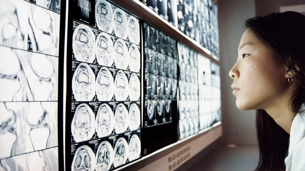
Some types of scans include X-rays, computed tomography (CT), magnetic resonance imaging (MRI), and ultrasound scans. Medical professionals use scans to help diagnose a range of health conditions.
They can also use them to track the progress of diseases, plan surgical procedures, and monitor pregnancies.
This article outlines what doctors use medical scans for. It also explores several common types of scans in more detail.

Medical scans help doctors
A doctor
- confirm a diagnosis
- rule out serious illnesses
- make a differential diagnosis
Medical professionals can also use scans to assist with surgical procedures. For example, they may carry out a scan
X-ray scans use X-rays to
During an X-ray scan, X-rays travel through the body. As they do this, different tissues in the body absorb different amounts of the rays.
The rays then pass through the body and into an X-ray detector on the other side of the body. The detector then creates an image that represents the “shadows” formed by the different tissues in the body.
Bones absorb large amounts of X-rays. This means that less of the rays pass through to the X-ray detector on the other side. This means that bones will appear whiter than other tissues in the image.
Tissues that X-rays pass through easily will then display in shades of gray. The more rays that pass through the tissue, the darker they will appear in the X-ray image.
Medical professionals can use X-ray scans to:
- locate bone fractures
- locate certain tumors
- diagnose pneumonia
- diagnose dental problems
- identify certain foreign bodies inside the body
- spot certain types of injuries
- locate abnormal masses in the body
- locate calcifications
CT scans are a type of imaging scan
During a CT scan, the radiographer will aim a narrow beam of X-rays at a specific part of the body. They then rotate the beams around the body. A digital X-ray detector then picks up these rays and creates a cross-sectional image of a part of the body. Medical professionals may refer to this image as a “slice”.
The CT machine’s computer may then digitally stack these slices on top of one another to create a three-dimensional image, which makes it easier for medical professionals to identify basic structures and spot abnormalities.
Medical professionals can use CT scans to:
- detect tumors in the:
- locate injuries in the brain
- locate blood clots in the brain that may lead to stroke
- locate hemorrhages in the brain
- detect heart diseases or abnormalities
- detect blood clots in the lungs
- detect excess fluid in the lungs
- diagnose lung conditions, such as emphysema and pneumonia
- create images of bone fractures and joint damage
Medical professionals use MRI scans to create detailed,
MRI scanners use
A radiologist will then use the MRI machine to pass radio waves through the body. These waves cause the protons in the body to move from their original position. When the radiologist turns the radio waves off, the protons will move back into their original position.
The MRI sensors can then detect the energy that these protons release as they realign with the magnetic field. The MRI computer then turns the energy that the MRI sensors detect into three-dimensional images.
MRI scans differ from CT scans and X-rays as they do not use the potentially damaging ionizing radiation of X-rays. The following structures appear more clearly in an MRI scan than regular X-rays or CT scans:
- brain
- spinal cord
- nerves
- muscles
- ligaments and tendons
MRI scans can help medical professionals to:
- locate tumors
- observe different brain structures
- determine which parts of the brain activate during certain tasks
- diagnose soft tissue injuries
- monitor the effects of certain treatments
- assess a person’s neurological status and neurosurgical risks
Ultrasound scans use ultrasound waves to
During an ultrasound scan, a medical professional will place a probe onto the person’s skin. This probe emits high-frequency ultrasound waves that bounce off certain parts of the body.
The ultrasound probe then picks up these waves as they bounce back. After this, the ultrasound waves hit the transducer and generate electrical signals, which then make their way to the ultrasound scanner. The scanner then turns these signals into a live, moving image of tissues or organs.
Medical professionals also use ultrasound scans to create images of organs and other parts of the body. They can use these to look for abnormalities in the:
Additionally, healthcare professionals use ultrasound scans to
- monitor the growth of the fetus
- carry out an anatomical survey of the fetus
- locate the placenta
- look at the amniotic fluid
- observe the pelvic organs in the pregnant person
An echocardiogram is
During an echocardiogram, a medical professional uses a small probe to send out high-frequency sound waves. These waves create echoes when they bounce off different structures in the body.
The probe then picks up these echoes and turns them into a moving image.
Echocardiograms
- the size and shape of the heart
- how well the heart is working
- problems with the heart valves
- weakness or dysfunction in the wall or muscle of the heart
- blood clots in the heart
- how a person is responding to medications and other treatments
An ECG is a type of test that medical professionals use
ECG tests can allow healthcare professionals to measure the time it takes for electrical waves to pass through the heart. ECGs also allow them to measure the amount of electrical activity that passes through the heart muscle.
This test can help medical professionals to understand whether the electrical activity in the heart is:
- fast
- slow
- irregular
It also allows them to determine:
- if there is damage to parts of the heart
- if parts of the heart are too large
- if parts of the heart are working too hard
ECGs can help medical professionals diagnose:
PET scans use radiation to create
During a PET scan, a medical professional will inject a person with a radiotracer. Alternatively, a person may swallow or inhale the radiotracer. This substance collects in different parts of the body.
The PET scanner can then detect the areas in the body where the radiotracer does and does not build up. The PET scanner then creates images of structures inside the body.
Healthcare professionals most commonly use PET scans to diagnose different types of cancer.
Medical professionals commonly use scans to help diagnose medical conditions.
They can also use scans to track the progress of diseases, monitor pregnancies, measure electrical activity in the body, and plan surgical procedures.
Common types of scans include X-rays, CT scans, MRI scans, ultrasound scans, echocardiograms, ECGs, and PET scans.


